Molecular Detection of Merozoite Surface Protein 1 and 2 in Plasmodium Falciparum Infected Subjects in Some Selected Hospitals, Bayelsa State, Nigeria
Promise Oboh Suama , Abigail Awudumaro Imeni , Nathaniel Festus Jack , Daniel Samuel1 ,
Department of Medical Laboratory Science, School of Allied Medical Sciences, College of Health Technology,
Otuogidi, Ogbia-Town, Bayelsa State, Nigeria.
Vivian Ibienebakabobo Promise2 ,
Department of Public Health, Faculty of HealthSciences, Bayelsa State Medical University
Yenagoa, Bayelsa State, Nigeria
Tolulope Alade3 , Oluwayemisi Agnes Olorode , Tatfeng Youtchou Mirabeau 3
33Department of Medical Laboratory Science, Faculty of Basic Medical Sciences, College of Health Sciences, Niger Delta University, Wilberforce Island, Yenagoa, Bayelsa State, Nigeria.
All Corresponding to: ohakpolamugwo Nwabueze C . Email:suamapro@gmail.com
Cell Phone: +23407064890721; Orcid: https://orcid.org/0000-0002-8288-3163
ABSTRACT
Abstrat: Malaria still remains a major burden, despite efforts in curbing this disease in Bayelsa state. To better manage and treat this disturbing disease, it is essential to detect merozoite surface protein 1 (msp1) and merozoite surface protein 2 (msp2) in Plasmodium falciparum infected subjects. This present study was aimed to detect msp 1 and 2 in Plasmodium falciparum infected subjects in Bayelsa state. Across sectional design was adopted. The parasite's DNA was extracted using kit method. A total of 50 positive malaria subjects with parasite density of ≥5,000/µl were selected for this study. However, samples with positive P.falciparum examined by microscopy were confirmed with the polymerase chain reaction technique. The msp1 and msp2 genes were amplified with Nested-Touch Dawn PCR method and their amplicons analyzed by electrophoresis on a 1% agarose gel stained with 5µl Safe-View dye. The alleles present were resolved according to their molecular ladder under a blue light. Forty percent (40.0%) of the positive samples were confirmed as P.falciparum by PCR. In msp1, RO33 and MAD20 alleles were detected of which, 10% RO33 alleles was predominant followed by 5.0% MAD20 alleles. In msp2, no alleles were observed. The association between multiplicity of infection and parasite density was -13 (P<0.05). This study detected only MSP1 in subjects being infected with P.falciparum. Drugs for the treatment of malaria should be design to target MSP1 strains of Plasmodium falciparum circulating in Bayelsa state.
Key words: Molecular Detection, Merozoite Surface Protein, Plasmodium falciparum, Infected Subjects, Malaria Parasitaemia
INTRODUCTION
Despite all the efforts by both governmental and non-governmental organizations to control malaria, it is still a global concern in respect to morbidity and mortality [1]. In line with the emergence of new strains of Plasmodia, Plasmodium strains resistant to Artemisinin Based Combination Therapy (ACT) emerging and the emergence of resistant vectors to insecticides is a major contributory factor to the endemicity of malaria parasites [2]. Five Plasmodium species are known to cause malaria in humans [3], such as the Plasmodium falciparum, Plasmodium malariae, Plasmodium vivax, Plasmodium ovale and Plasmodium knowlesi [4]-[6].
Malaria, a tropical disease is caused by Plasmodium species and mainly acquired through the bite of an infected female anopheles mosquito [7], [8]. Among all the Plasmodium species, Plasmodium falciparum is the deadliest, caused from mild to severe malaria conditions associated with morbidity and mortality. Additionally, it is frequently accompanied with antimalarial drug resistance and failed vaccines trials [9].
It had been reported that genetic variety of Plasmodium falciparum plays an imperative role in defining the power in the spread of malaria. However, numerous Plasmodium falciparum genes had shown extensive genetic polymorphism in merozoite surface proteins 1 (msp1) and merozoite surface proteins 2 (msp2) in different
To overcome its isolation challenge, a modification of TB growth medium will be considered in this research to improve the growth rate and identification process.. The aim of this research is therefore to improve L-J medium to facilitatefaster growth of MTB, identify the Mycobacterium tuberculosis complex (MTBC) and determine the rifampicin resistant MTB isolates. environmental sites where malaria is endemic. In addition, msp1 and msp2 genes are extensively used genes to study the allelic multiplicity and incidence of Plasmodium falciparum [10]. These genetic markers are essential molecular tool used in detecting malaria parasites [11], [12]. However, merozoite surface protein 2 (MSP2) plays some vital roles as major vaccine candidates while merozoite surface protein 1 (MSP1) facilitates the invasion of red blood cells in the human host [13].
It is commonly reported that diagnosis of malaria is routinely examine with Giemsa stained blood film under light microscope [3]. However, malaria parasites (Plasmodium falciparum for instance) is better and more efficiently detected with the polymerase chain reaction (PCR) by amplifying the genes coding for msp1 and msp2 [14]. Therefore, the interest in this present study is geared towards detecting msp1 and msp2 in Plasmodium falciparum infected subjects in some selected hospitals in Bayelsa state.
MATERIALS AND METHODS
Study Design
A cross-sectional observational study design was adopted. A cross-sectional observational study design was on Plasmodium infected subjects with malaria parasites density ≥ 5000 parasites/μl of blood at Molecular Laboratory Unit in the Medical Laboratory Science Department, Niger Delta University, Bayelsa state.
Study Site
The study was carried out in Bayelsa state, Nigeria. There are eight local government in Bayelsa state, with its capital situated in Yenagoa whereas the state is divided into three federal senatorial districts. This senatorial district includes: The Central that comprising of Yenagoa, KulokumoOpukuma and Southern Ijaw Local Government Areas, West senatorial district comprising of Brass, Nembe and Ogbia Local Government Areas, while East senatorial district comprising of Sagbama and Ekeremor Local Government Areas respectively as shown in [figure 1]. Accordingly, the 2006 census stated that Bayelsa state has a population of approximately 1.7million people. It is o o geographically located latitude 4 151north, 5 231south and o o longitude 5 221west, 6 451east. Also, bounded with Delta state on the north, Rivers state on the east and the Atlantic Ocean on the west and south [16].

Study population:
The population of this study included all subjects that were diagnosed with Plasmodium falciparum malaria by microscopy and haven parasitaemia levels of ≥ 5000 parasites/μl in the selected hospitals in Bayelsa state. Inclusion criteria: The followings were the inclusion criteria used for this work.
- Subjects from the ages of six (6) months old visiting the laboratory with fever or physician clinical diagnosis of malaria
- Subjects from the ages of six (6) months old visiting the laboratory with complaint of fever
- Subjects from the ages of six (6) months old visiting the laboratory with absence of documented malaria treatment in the past two weeks prior to this work
- Only subjects confirmed infected with Plasmodium falciparum with parasite density of ≥5,000 parasites/μl from examination of Giemsa stained blood film were used for molecular analysis.
Exclusion criteria:
The exclusion criteria for this present study were:
- Subjects from ages of six (6) months old visiting the laboratory who refuged granting consent
- Subjects from ages of six (6) months old visiting the laboratory noted of having evidence of any documented adverse drug effect.
Malaria Parasite Detection
Thick and thin blood films were prepared, stained with a 10% Giemsa working solution and examined with ×100 objective lens under a light microscope. Parasites detected were counted simultaneously with differential white blood cells, and parasites density were calculated using the formula given below

DNAExtraction
Parasites DNA were extracted using the kit extraction method. Approximately, 400µl of genomic lysis was added to 100µl of blood, mixed, vortexed for 5 seconds and incubated at room temperature for 10 minutes. The mixture was transferred to a Zymo-Spin IIC column in a collection tube and centrifuged at 10000x g for 1 minute. The collection tube along with their flow through were discarded. The Zymo-Spin IIC column of the mixture was transferred to a new collection tube. A 200µl DNA Prewash Buffer was added to the spin column and centrifuged at 10 000xg for 1muinute. A s500µl of g-DNA wash buffer was added to the spin column, centrifuged again at 10000x-g for 1minute. The spun column was transferred into another clean microcentrifuge tube, 50µl of DNAelution buffer was added to the column, incubated at room temperature for 5minutes and centrifuged at top speed for 30seconds to elute the DNA. The eluted DNAwas tested for its purity, quantity o o of the content and stored at 5 C-20 C for further analysis.
DNAPurification and Quantification
The purity and the quantity of the DNAwas measured using Nano-drop 1000 Spectrophotometer. Using software appt, it was lunched by clicking the Nano-drop icon twice while the equipment was initialized with 2µl of sterile distilled water and blanked with normal saline. 2µl of each extracted DNA was loaded separately onto lower pedestal, the upper pedestal was brought down to make contact with the extracted DNA placed on the lower pedestal. The quantity and purity of the DNA were read at interval with a corresponding software. The ratio of absorbance (260/280) was used to determine the DNA purity as previously adopted [17] - [18], [9]
Molecular Detection of Plasmodium falciparum
Polymerase Chain Reaction was used to confirm malaria positive samples examined with the microscopic Giemsa stained blood films. P.falciparum genes as shown in (Table i) was used. The reaction was performed in a final volume of 30μl containing: 2X Master-Mix, water, extracted P.falciparum DNA template, forward and reverse primers according to the manufacturer's instructions.
The reaction was performed with 30μl containing 2X Master-Mix and water, amplicon from the first PVR product as DNA template, forward and reverse primers. In these reactions, 1μl each of the first PCR product were added as the DNA templates. Each amplification conditions were set o o as follows: For msp1, it was set at 98 C for 5min, 95 C for o o o o 40s,75 C for 1min, 70 C for 1min, 68 C for 40secs, 69 C for o 5mins and a holding period at 72 C for 10mins in 30 cycles o o o while msp2 condition was 98 C for 5min, 95 C for 40s, 75 C o o o for 1min, 72 C for 1min, 68 C for 40secs, 69 C for 5mins o and a holding period at 72 C for 10mins in 30 cycles respectively. While their PCR results were resolved using agarose gel electrophoresis [19]. The prevalence of each

PCR Amplification: In this reaction, a 2μl of the extracted DNA was added to the PCR component. The PCR condition was o o o o o o o set at 98 C for 5min, 95 C for 40s,75 C for 1min, 70 C for 1min, 68 C for 40secs, 69 C for 5mins and a holding period at 72 C for 10mins in 30 cycles. The result PCR was resolved using agarose gel electrophoresis [19]. A nested touch down PCR technique was used to detect the P.falciparum msp1, P.falciparum msp2 genes. Specific primers designed as shown in (Table ii) were used to amplify msp1 and msp2 genes in the primary reactions.
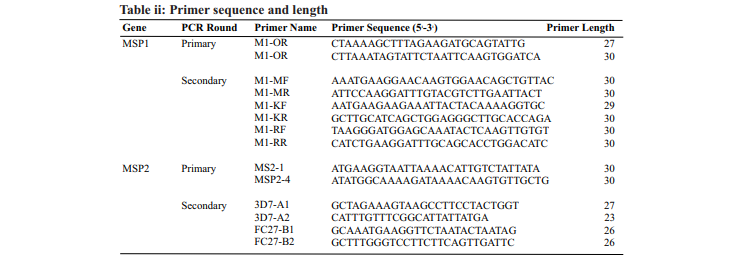
allelic type was determined as the presence of PCR products for the type in the total number of amplified bands for the corresponding locus. A nested touch down PCR technique was used to detect the P.falciparum MSP1, P.falciparum MSP2 genes. Specific primers designed as shown in (Table ii) were used to amplify the alleleic families in the secondary reactions. The reactions were performed in a final volume of 30μl containing: 2X Master-Mix, water, amplicon from the first PVR product as DNA template, forward and reverse primers. In these secondary reactions, 1μl each of the first PCR product were added as DNA templates according to the manufacturer instructions. In msp1, each amplification conditions were set as follows: the o o MAD20 families were set at 98 C for 5min, 95 C for 40s, o o o o 83 C for 1min, 78 C for 1min, 68 C for 40secs, 69 C for o 5mins and a holding period at 72 C for 10mins in 30 cycles; o o o K1 allelic families were at 98 C for 5min, 95 C for 40s, 86 C o o o for 1min, 81 C for 1min, 68 C for 40secs, 69 C for 5mins o and a holding period at 72 C for 10mins in 30 cycles and the o o RO33 allelic families were set at 98 C for 5min, 95 C for o o o o 40s, 86 C for 1min, 81 C for 1min, 68 C for 40secs, 69 C for o 5mins and a holding period at 72 C for 10mins in 30 cycles [19]. In msp2, each amplification conditions were set as o follows: the 3D7 allelic families were set at 98 C for 5min, o o o o 95 C for 40s, 70 C for 1min, 65 C for 1min, 68 C for 40secs, o o 69 C for 5mins and a holding period at 72 C for 10mins in 30 o o cycles and FC27 allelic families were at 98 C for 5min, 95 C o o o o for 40s, 72 C for 1min, 67 C for 1min, 68 C for 40secs, 69 C o for 5mins and a holding period at 72 C for 10mins in 30 cycles [19].
DNAElectrophoresis
The PCR products were resolved and sized against a 100bp molecular weight marker on a 1% agarose gel made in a 1X Tri-Boris EDTA containing 5µl Safe-View and ran in a horizontal tank filled with 1X Tri-Boris EDTA for 30mins at 350V. All gels were visualized under a blue light transillumination and the electrophoretic condition was 100mV for 30min. The size and fragment of the respective bands were estimated by comparing bands with a 100kb plus DNAladder [19].
Multiplicity of Infection
The Multiplicity of Infection (MOI) or number of genotypes per infection was calculated by dividing the total number of fragments detected in msp1 and msp2 by the number of samples positive for the same marker [9]. Polyclonal infections were more in majority of the patients for msp1 allelic families [20].
Data Analysis
All data obtained from the results of the Giemsa stained blood films were entered into Statistical Package for the Social Science (SPSS) version 23 and analyzed using analyzed using descriptive statistics for the categorical variables, while an inferential was used for continuous variables. The allelic frequency of msp1 and msp2 was calculated as proportion of the allele detected for each of the allelic family from the overall total alleles detected. The frequency of polyclonal infection was calculated using number of samples with more than one amplified fragment out of the total samples. Multiplicity of infection was determined by dividing the total number of alleles detected in msp1 and msp2 by the total number of positive samples for both genetic marker. Descriptive statistics was used to calculated mean standard deviation of the malaria parasitaemia, and results were expressed in percentages and presented in frequencies. While Spearman rank correlation coefficient (r), was used to determine the probable associations exiting between the parasitaemia level and multiplicity of infection. Statistical significance was defined as p ≤0.05
RESULTS
Table 1a showed the distribution of malaria parasitaemia among the Plasmodium falciparum infected subjects in some selected hospitals Bayelsa state, and level of parasitaemia varied between < 5000 parasites/µl to ≥ 15,000 parasites/µl of blood. Of the 180 positive cases, 130(72.2%) had malaria parasitaemia less than 5000 parasites/µl of blood and 50(27.8%) had malaria parasitaemia of 5000 parasites/µl of blood and above. Out of the 50 subjects with higher malaria parasitaemia density, 22(12.2%) had parasitaemia of 5000-10000 parasites/µl of blood, 21(11.7%) had parasitaemia of 10001-15000 parasites/µl of blood and 7(3.9%) had parasitaemia above 15000 parasites/µl of blood
Prevalence of malaria parasitaemia based on gender of subjects were 65.9% of which, 72.2% had malaria parasitaemia of >5000 parasite/µl (consisting 62 males and 68 females) while 50(27.8%) subjects malaria parasitaemia of ≥ 5000 parasite/µl. Of this, 16.7% of subjects had parasitaemia level between 5000-10000 parasite/µl of which, 12% males and 1.8% females, ffollowed by 8.3% of subjects had parasitaemia level between 10001-15000 parasite/µl of which, 6% males and 10% females and 16.7% of subjects had highest parasitaemia level of >15000 parasite/µl of which, 7.2% males and 8.6% females respectively.
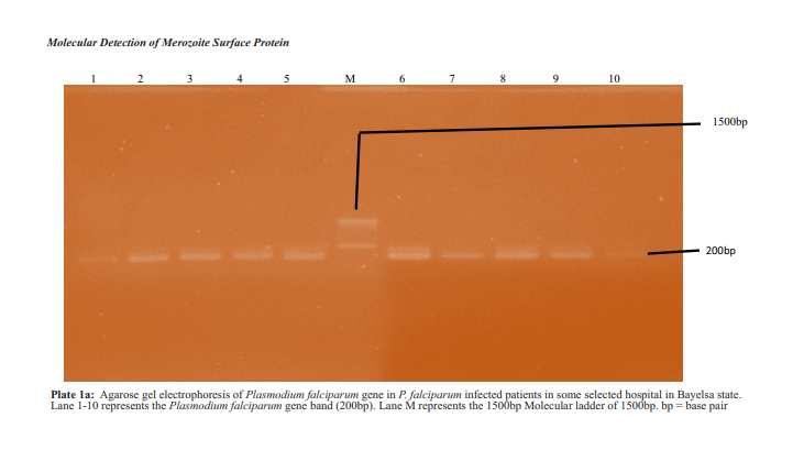
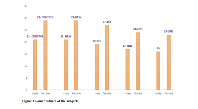
Table 2 and plate 1a showed PCR distribution of Plasmodium falciparum by Age and Gender. Parasite genomic DNA was extracted from the 50 blood samples with the highest parasitaemia levels ≥5000 parasites/µl of blood of which, 20(40.00%) P.falciparum were confirmed by PCR. Of this number, 8(38.1%) were from males while 12(41.4%) females. In males: eight (8) malaria parasites were examined from patients below 11 years old of which, 3(37.5%) P.falciparum were confirmed. Five (5) malaria

The prevalence of malaria parasitaemia based on age of the subjects were 65.9% of which, 130(72.2%) of subjects had malaria parasitaemia of >5000 parasite/µl while 50(27.8%) subjects malaria parasitaemia of ≥ 5000 parasite/µl.
Table 1b showed the distribution of malaria parasitaemia (parasite density) among Plasmodium falciparum infected patients by gender in some selected hospitals Bayelsa state. Of the 180 malaria positive patients, 130(72.2%) had parasite density of less than 5000 parasites/µl of blood while 50(27.8%) had parasite density of 5000 and above parasites/µl of blood. Of the 50 malaria positive patients with parasite density of 5000 and above parasites/µl of blood, 21(16.7%) had parasite density between 5000-10000 parasites/µl of blood, 15(8.3%) had parasite density between 10001-15000 parasites/µl of blood and14(7.8%) had parasite density above 15000 parasites/µl of blood.

parasites were examined from patients between the ages of 11-20 years of which, 2(40.0%) P.falciparum were confirmed. Three (3) malaria parasites examined from patients between the ages of 31-40 years of which, 1(33.3%) P.falciparum were confirmed and four (4) malaria parasites were examined from patients between the ages of 41-50 years of which, 2(50.0%) P.falciparum were confirmed. In females: nine (9) malaria parasites were examined from patients below 11 years old of which, 7(77.8%) P.falciparum were confirmed. Two (2) malaria parasites were examined from patients between the ages of 11-20 years of which, 1(50.0%) P.falciparum were confirmed. Six (6) malaria parasites examined from patients between the ages of 31-40 years of which, 1(16.7%) P.falciparum were confirmed. Five (5) malaria parasites were examined from patients between the ages of 41-50 years of which, 2(40.0%) P.falciparum were confirmed and two (2) malaria parasites were examined from patients over the ages of 70 years of which, 1(50.0%) P.falciparum were confirmed respectively
Discussion Of the 50 samples taken for PCR with malaria parasitaemia ≥ 5000/µl of blood, 20(40.00%) showed amplification while 30(60.0%) showed no amplification for P.falciparum respectively. Reason could possibly be due to specificity and sensitivity of the choice of primers used. This finding was in agreement with other study [9], but lower in similar studies [21]. This finding is also in conformity with a previously reported case of importance of molecular genotyping of P.falciparum [14]. However, genotyping of Plasmodium falciparum msp1 and msp2 are recommended for the antimalaria clinical trials as standard markers to distinguish recrudescent from a newly infecting malaria episode [22]. Also, to the best of our knowledge, this kind of study investigating the genetic diversity of msp1 and msp2 in malaria parasites circulating hospitals in Bayelsa state. Hence, our study aimed to detect msp1 and msp2 among Plasmodium falciparum infected subjects in hospitals, Bayelsa state is scarcity
pitals, Bayelsa state is scarcity. Successfully, this study detects only the msp1 gene in Plasmodium falciparum infected subjects. This might suggest that only this strain of the parasite seems to be

Key: (NE) = Number Examined, (NI) = Number Infected, (%) = Percentage, < = Less than and > = Greater than
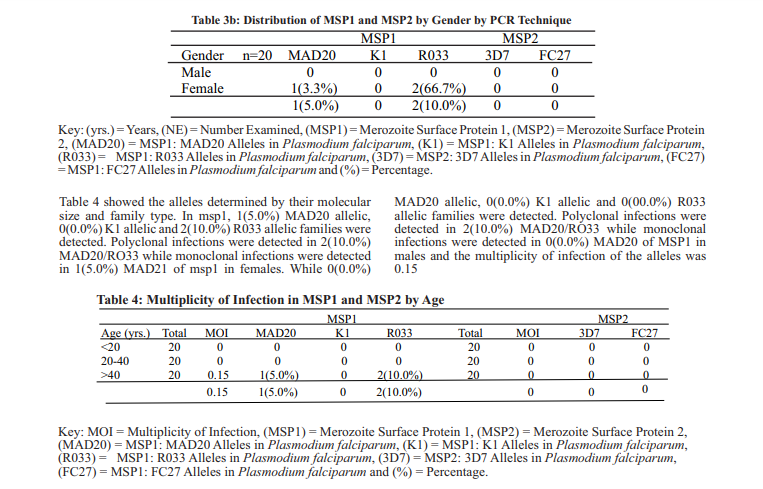
Of the 20 P.falciparum from patients were detected for msp1 and msp2 by age using nested touch down PCR, 2(10.0%) msp1 and 0(0.0%) msp2 were detected in patients within the aged of 40 years and above. In msp1, 1(20.0%) MAD20 allelic, 0(0.0%) K1 allelic and 2(40.0%) R033 allelic families were detected. Polyclonal infections were detected in 2(10.0%) MAD20/RO33 while monoclonal infections were detected in 1(5.0%) MAD21 of msp1 respectively. In MSP2, 0(0.0%) 3D7 allelic and 0(0.0%) FC27 allelic families were detected. Polyclonal infections were detected in 0(0.0%) and monoclonal infections were detected in 0(0.0%) of the allelic families respectively as shown in (Table 3a).

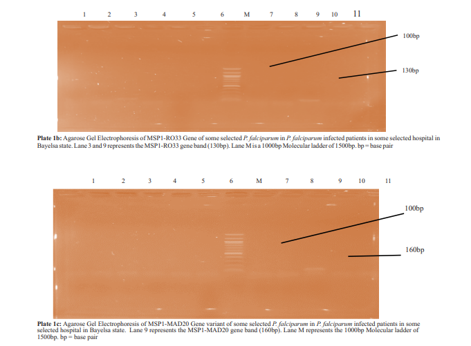
A total of 20 P.falciparum from patients were detected for msp1 and msp2 by gender using nested touch down PCR of which , 2(10.0%) msp1 genes and 0(0.0%) msp2 genes were detected. For MSP1, 0(0.0%)MAD20, 0(0.0%)K1 and 0(0.0%)R033 alleles were detected in males while 1(5.0%)MAD20 alleles, 0(0.0%)K1 alleles and 2(40.0%)R033 alleles in females. A polyclonal infection was detected as 2(10.0%)MAD20/RO33 and monoclonal infection as 1(5.0%)MAD21 in MSP1. For MSP2, 0(0.0%)3D7 and 0(0.0%)FC27 alleles were detected in males and females respectively as shown in (Table 3b and Plate 1b-f).
In total, 15.0%%(2/20) and 0% of the parasite strains contained multiplicity of infections (MOIs) on msp1 and msp2 respectively. The MOIs for both msp1 and msp2 are summarized in table 5. The MOI for msp1 was 0.15 with a range of 1-2 strains and was higher than MOI for msp2 of 0 with zero range of strains. There is no significance association existing between malaria parasitaemia and multiplicity of infections in P.falciparum infected subject in hospitals in Bayelsa state. The Spearman's correlation coefficient (r) was - 0.13 at a mean ± SD of 3112.63 ± 10256.00 parasites/μl of blood (p = 0.37) as in table 8. The mean ± SD of malaria parasitaemia was 3112.63 ± 10256.00 and multiplicity of P.falciparum infection was 0.15, association (r) between malaria parasitaemia and multiplicity of infection was -0.13 at P-vale of 0.37

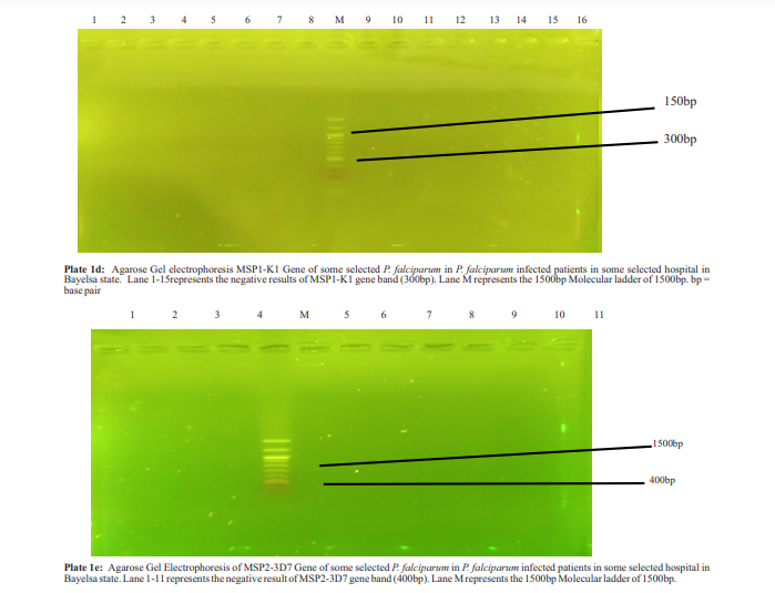
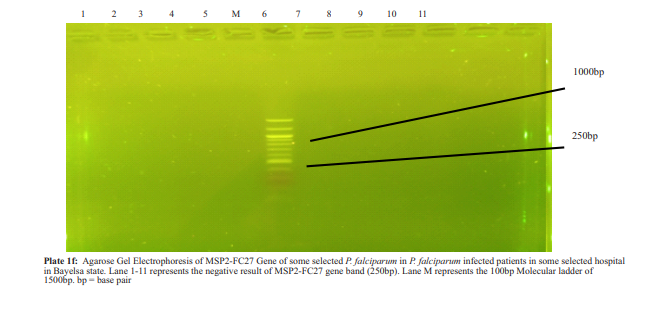
predominant in the state. This finding conformed but slightly differ with the previous studies done [9]. Genetic polymorphism the merozoite surface proteins of Plasmodium falciparum detected were analyzed demonstrated allelic specific PCR in the genotyping msp1 showed the presence of R033 and MAD20 allelic families in the Plasmodium falciparum. Of the two allelic families of msp1, R033 was the most predominant, followed by MAD20 allelic family as it was identified. This finding revealed the pattern of distribution and suggestive of the malaria parasites strains circulating in Bayelsa state. It added to the existing knowledge of the P.falciparum strains predominate in Bayelsa state. Also, not understanding that P.falciparum with MAD20 and R033 alleles are the strains infecting subjects might be the reasons for poor management recurrent malaria in the state. This finding, pattern of allelic distribution is in agreement with patterns observed [9], [10], [23], but inconsistent with [21]
In this study, P.falciparum haven the msp2 stain and the FC27 and 3D7 allelic families were not detected. This might be that P.falciparum possessing this strain is not predominant in Bayelsa state. Although, this finding appeared to be inconsistent with previous study done [21]. The multiplicity of infection observed in this study was highest for msp1 (0.15) and lowest for msp2 (0.00) when compared with values from other works done elsewhere. Probably this might be why malaria is still very poorly treated in Bayelsa state hence, making this disease endemic in the state. This finding was contrary to other work done [10], This finding also showed that the association between the multiplicity of infection and parasitaemia levels in the P.falciparum infected subjects in selected hospitals in Bayelsa state is statistically significant.
CONCLUSION
These results demonstrated high genetic diversity only in msp1 strain of Plasmodium falciparum with the MAD20 and R033 alleles. Therefore, it had imperative that drugs design in treating malaria in Bayelsa state should be targeted towards the msp1 strains of Plasmodium falciparum
ACKNOWLEDGEMENT
Mr Ishmael Ebidimie- (General University Hospital, Sagbama), Mrs Nake Nelson. (Federal Medical Centre Otuoke) and Mrs Martha Ukpong (Federal Medical Centre Yenagoa (They greatly assisted in sample collection)
REFERENCES
- World Health Organization. World malaria report 2016. Accessed at rd https//: www.who.int.mal-publications. on 3 March 2020
- World Health Organization. World malaria report 2012. Accessed at https://www.who.int/malaria on16th February 2023
- Caraballo, H., & King, K. (2014). Emergency department management of mosquito-borne illness: malaria, dengue, and West N i l e virus. Emergency Medicine Practice. 16 (5) 1–23
- Erhabor, O., Azuonwu, P., & Frank-Peterside, N. (2012). Malaria parasitaemia among long distance truck drivers in the Niger Delta of Nigeria. African Health Sciences. 12(2), 98-103.
- Adeyemo, O., Okpala, U., Nwachukwu, E., & Imoukhuede, O. (2014). Malaria infection amongst students of the university of Benin, Edo State, Nigeria. International Journal of Recent Scientific Research. 5(9), 1529-1532.
- Butcher, A., & Mitchell, H. (2016). The role of Plasmodium knowlesi in the history of malaria research. Parasitology. 145(1), 6- 17.
- ), 6- 17. 7. Centre for Disease Control and Prevention. Treatment of malaria parasitemia in pregnant women. Accessed at https//: www.cdc.gov. st mal.impact on 1 February 2020
- World Health Organization. World malaria report 2017. Accessed at th https//: www.who.int.mal-publications on 6 June 2021
- Olatunji, K., Olugbenga, M., Yetunde, O., & Tolulope, O. (2016). Population genomics diversity of Plasmodium falciparum in malaria patients attending Okelele Health Centre, Okelele, Ilorin, Kwara State, Nigeria. African Health Science. 16(3), 704-711.
- Mwingira, F., Nkwengulila, G., Schoepflin, S., Sumari, D., Beck, P., Snounou, G., Felger, I., Olliaro, P., & Mugittu, K. (2011). Plasmodium falciparummsp1, msp2 and glurp allele frequency and diversity in sub-Saharan Africa. Malaria Journal. 10, 79.
- Manikande, B. (2012). Genetic mapping and marker assisted selection: Basic practice and benefits. Springer science and business media, United State of America. PP. 60-81
- Ghansah, A., Amenga-Etego, L., & Amambua-Ngwa, A. (2014). Monitoring parasite diversity for malaria elimination in subSaharan Africa. Science. 345(6202), 1297–1298.
- Beeson, G., Drew, R., Boyle, J., Feng, G., Fowkes, J., & Richards, S. (2016). Merozoite surface proteins in red blood cell invasion, immunity and vaccines against malaria. Federation of European Microbiological Societies and Microbiology Reviews. 40 (3), 343–372
- Bakhiet, M., Abdel-Muhsin, M., Elzaki, E., Al-Hashami, Z., Alberwani, S., & Al-Qamashoul, A. (2015). Plasmodium falciparum population structure in sudan post artermesinin-based combination therapy. Acta Tropica. 148, 97-104
- Brisibe, W.., & Brown, I. (2020). Map of Bayelsa state. International Journal of Architectural Engineering Technology. 7(4), 36-46
- Northgate Exploration Limited. Bayelsa state, Nigeria. Accessed at rd https://www.ngex.como 3 July 2022
- Desjardins, R., & Conklin, S. (2011). Microvolume quantitation of nucleic acid. Current Protocols in Human Genetics. 93(1), 1–16.
- Sukumaran, S. (2013). Concentration determination of nucleic acids and proteins using the micro-volume Bio-Spec Nano Spectrophotometer. Journal of Visualized Experiments. 15(48), 2699
- Haanshuus, G., Mohn, C., & Morch, K. (2013). A novel, singleamplification PCR targeting mitochondrial genome highly sensitive and specific in diagnosing malaria among returned travelers in Bergen, Norway. Malaria Journal. 12, 26.
- Atroosh, M., Al-Mekhlafi, M., Mahdy, K., Saif-Ali, R., AlMekhlafi, M., & Surin, J. (2011). Genetic diversity of Plasmodium falciparum isolates from Pahang, Malaysia based on msp-1 and msp-2 genes. Parasites and Vectors. 4, 233
- Somé, F., Bazié, T., Zongo, I., Yerbanga, S., & Nikiéma, F. (2018). Plasmodium falciparummsp1 and msp2 genetic diversity and allele frequencies in parasites isolated from symptomatic malaria patients in Bobo-Dioulasso, Burkina Faso. Parasitological Vectors. 11, 323-330.
- Ferrar, T., Deperalta, K., Day, H., Rietz, A., Wei, L., & Lauwers, Y. (2015). 3-D molecular MR imaging of liver fibrosis and response to rapamycin therapy in a bile duct ligation rat model. Journal of Hepatology. 63, 689-696.
- Mohammed, H., Mindaye, T., Bilayneh, M., Kassa, M., Assefa, A.., & Tadesse, M. (2015). Genetic diversity of Plasmodium falciparum isolates based on msp1 and msp2 genes from KollaShele area, Arbaminch Zuria district, South-West Ethiopia. Malaria Journal. 14, 73


Comments are closed.